
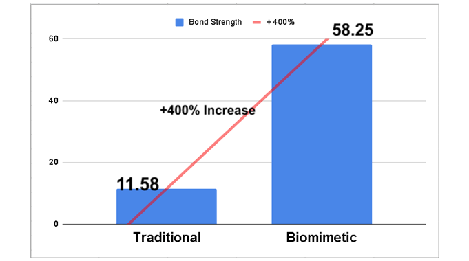
About a year ago, I was looking for background information for a research project analyzing the information shared about biomimetic restorative dentistry on social media, particularly Instagram. During that process, I came across several claims that caught my attention. One of them stated that biomimetic protocols can achieve bond strength values more than 400% higher than those obtained with conventional dental protocols.
The numbers were striking: 58.25 MPa vs. 11.58 MPa. I immediately suspected that they referred to a procedure known as immediate dentin sealing. Because of the amount of information involved, I have divided this post into two parts. In this first part, I will discuss the technique and its potential effect on dentin bond strength.
What is immediate dentin sealing (IDS)?
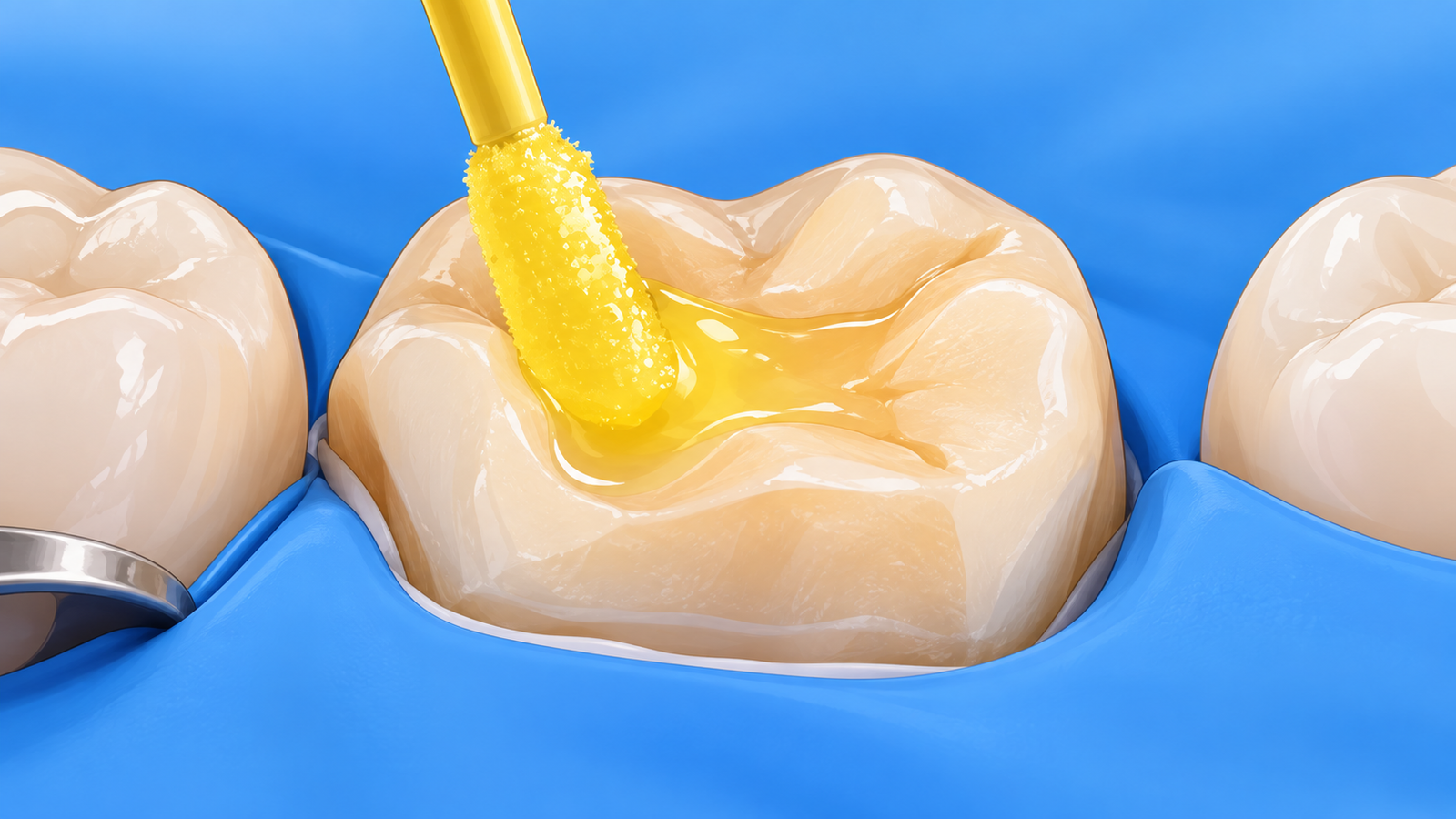
IDS is a procedure in which an adhesive system is applied and light-cured on freshly exposed dentin immediately after tooth preparation for an indirect restoration, before the impression is made and the provisional restoration is placed. The concept has its roots in strategies developed between the 1990s and early 2000s, such as the resin coating technique in Japan, dual bonding in Switzerland, and prehybridization in the United States. Pascal Magne later consolidated and popularized the concept of IDS.
Its potential advantages include protecting dentin during the provisional phase, reducing postoperative sensitivity, lowering the risk of contamination of the dentin surface, and allowing the adhesive interface to be fully polymerized before the definitive restoration is bonded or placed.
Before IDS was introduced, the conventional approach was delayed dentin sealing (DDS). In this sequence, the tooth was prepared, an impression was made, a provisional restoration was placed, and the adhesive procedure was performed only at the appointment for bonding the definitive indirect restoration.
When it comes specifically to improving bond strength, the claim of an increase greater than 400% did not come out of nowhere. It essentially originates from a classic study published by Magne et al. in 2005. In that experiment, bond strength was 11.58 MPa with DDS and 58.25 MPa with IDS, an impressive difference. But does simply waiting a couple of weeks before bonding really cause dentin to lose such a large proportion of its bonding potential?
The classic study by Pascal Magne et al. (2005) on IDS
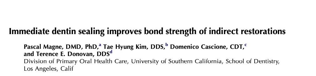
In December 2005, Magne et al. published an article in The Journal of Prosthetic Dentistry titled Immediate dentin sealing improves bond strength of indirect restorations. As of September 2026, it is the third most cited publication in Pascal Magne’s body of work, with 220 citations in Scopus and 514 in Google Scholar.
The authors used 15 human molars divided into three groups (n = 5):
- Control group: 1. Prepared dentin → 2. acid etching/primer/bonding agent (light-cured) → 3. direct incremental composite restoration
- DDS group: 1. Prepared dentin → 2. provisional restoration (2 weeks in saline solution) → 3. provisional removal and airborne-particle abrasion of dentin → 4. acid etching/primer/bonding agent (not light-cured) → 5. direct incremental composite restoration
- IDS group: 1. Prepared dentin → 2. acid etching/primer/bonding agent (light-cured) → 3. provisional restoration (2 weeks in saline solution) → 4. provisional removal and airborne-particle abrasion of the adhesive surface → 5. application of bonding resin only (not light-cured) → 6. direct incremental composite restoration
The study used the three-step etch-and-rinse adhesive OptiBond FL (Kerr) and Z100 composite resin (3M ESPE). Interestingly, although the title refers to improved bonding of indirect restorations, the experimental model simulated the provisional phase but did not include an indirect restoration or a resin cement. The final restoration was built directly using incrementally placed composite resin.
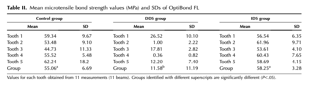
The results were striking: 55.06 MPa for the control group, 11.58 MPa for DDS, and 58.25 MPa for IDS. There was no statistically significant difference between the control and IDS groups, whereas DDS showed significantly lower bond strength. The authors also noted that the very large variability within the DDS group was caused by numerous failures occurring at loads close to zero. At first glance, the study appears to show that bonding to freshly prepared dentin is clearly superior to bonding two weeks later.
The failure pattern was also very different. In the DDS group, 98% of failures were interfacial, compared with 49% in the IDS group. In addition, although a 3–5 µm hybrid layer formed in the DDS specimens, the authors frequently observed discontinuities between this layer and the overlying resin. This is important because it suggests that the weakness was not necessarily related to adhesive penetration or hybrid layer formation itself, but may instead have been located in a more superficial region of the interface.
It is also important to clarify that the OptiBond FL values for the control, DDS, and 2-week IDS groups originally published in 2005 were reused in the 2007 article, which included additional experimental groups. Therefore, for evidence-synthesis purposes, these groups should not be considered independent findings from two separate investigations, because the data published in 2005 were included again in the 2007 paper.
With results showing such a dramatic difference, both the article and the IDS technique quickly gained attention. However, later studies by other research groups, which received considerably less attention than Magne’s work, did not necessarily reproduce the same pattern in favor of IDS.
Other studies with different results
Several studies have investigated the effect of IDS on bonding, but not all of them used methodologies similar enough to the classic 2005 Magne study to allow direct comparison. Some introduced additional variables, such as resin cements and indirect restorative materials, to simulate adhesive luting procedures. Examples include Rigos et al. (2019), Sakr et al. (2021), and Gailani et al. (2021).
However, among the published studies, two conducted by different research groups used methodologies that were particularly similar to that of Magne et al. (2005) and were published in internationally visible journals: Ribeiro da Silva et al. (2016) and van den Breemer et al. (2019).
The South American study on IDS
In 2016, Ribeiro da Silva et al., from the University of North Paraná in Londrina, Brazil, published an interesting study in Applied Adhesion Science examining the effects of DDS and IDS on bonding using another three-step etch-and-rinse adhesive: Adper Scotchbond Multi-Purpose (3M ESPE).
The article received considerably less attention than the Magne study. It is difficult to know why. Factors may include the fact that it was published in a journal outside the specific field of dentistry and that its results did not reveal a dramatic difference between IDS and DDS. As a reference, as of September 2026, the Ribeiro da Silva study has only 12 citations in Google Scholar, compared with 514 for Magne’s article.
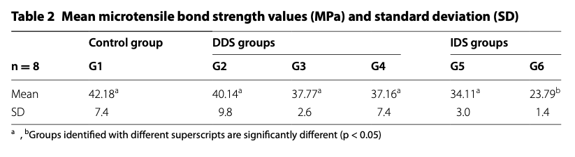
The control group (G1) was bonded immediately, as in a conventional direct restoration, and achieved 42.18 MPa. In one of the DDS groups (G4), the procedure was quite similar to that used by Magne et al. (2005). The dentin remained under a resin-based provisional material, Clip F, for two weeks in saline solution. The provisional material was then removed, the surface was cleaned first with a curette and then with pumice, and the adhesive procedure was performed before placement of Z350 XT composite resin. This group achieved 37.16 MPa, with no statistically significant difference from the control. The IDS group (G5), in turn, achieved 34.11 MPa. In fact, there were no statistically significant differences among the control, DDS, and IDS groups. In other words, after two weeks of provisionalization, the dramatic deterioration reported by Magne was not observed, even though the study used the same general type of resin-based provisional material.
Failure-mode analysis also did not show a clearly unfavorable pattern for DDS. Mixed failures predominated in both DDS and IDS (52.0% and 60.4%, respectively), whereas adhesive failures accounted for 18.8% and 22.9%.
It is also worth mentioning that groups G2 and G3, in which acrylic provisional restorations were cemented with Dycal (calcium hydroxide cement)) and TempBond NE (zinc oxide non-eugenol based cement), respectively, also showed no statistically significant differences compared with the control group, with values of 40.14 and 37.77 MPa.
The European study on IDS
In 2019, Carline van den Breemer from the University of Groningen in the Netherlands, together with other collaborators, published an experiment using a similar methodology. One important methodological difference was that OptiBond FL Adhesive was heated to 40 °C in all groups. Overall, the teeth remained for three weeks with a Protemp 4 bis-acryl provisional restoration, cemented with Durelon, a zinc polycarboxylate cement.
After removal of the provisional restoration, when the dentin in the DDS group was cleaned only with pumice, bond strength was 30.2 MPa, virtually identical to the 29.8 MPa obtained with a single-layer IDS strategy, with no statistically significant difference. In other words, even after three weeks of provisionalization, DDS and IDS did not differ significantly under this protocol.
In the DDS group, all evaluated failures occurred at the adhesive-dentin interface, and one pretest failure was also recorded. In the IDS group, 67% of failures were interfacial, 22% occurred within the adhesive, and 11% occurred within dentin. However, the number of specimens was small, and the study did not report a statistical comparison of failure-mode distributions. These differences should therefore be interpreted descriptively only.
To facilitate comparison with Magne et al. (2005), I selected only the results obtained after 1 week for the single-layer IDS group (IDS-1L) and excluded the groups involving two adhesive layers, an additional layer of flowable composite, and the results obtained after 6 months of storage. I also excluded the groups in which pumice cleaning was followed by tribochemical airborne-particle abrasion with CoJet, using silica-coated aluminum oxide particles, because adding these variables would make comparison among the studies more difficult. This analysis is not intended to have the methodological rigor of a systematic review. Rather, its purpose is to identify findings in the literature showing that DDS does not always result in a significant reduction in bond strength compared with IDS.
Comparison of the three studies
To summarize, I prepared the following table comparing the main methodological characteristics of these three studies and highlighting their similarities:
| Characteristic | Magne et al. 2005 | Ribeiro da Silva et al. 2016 | van den Breemer et al. 2019 |
|---|---|---|---|
| Strategy compared | DDS vs. IDS | DDS vs. IDS | DDS vs. IDS |
| Group designation in the study | DDS and IDS | DDS (G4) and IDS (G5) | DDS (SC-P/1 wk) and IDS-1L (SC-P/1 wk) |
| Substrate | Human molars | Human molars | Human molars |
| Surface | Flat coronal dentin | Flat coronal dentin | Flat coronal dentin |
| Adhesive | OptiBond FL | Adper Scotchbond Multi-Purpose | OptiBond FL |
| Adhesive type | 3-step etch-and-rinse | 3-step etch-and-rinse | 3-step etch-and-rinse |
| Provisionalization period | 2 weeks | 2 weeks | 3 weeks |
| Storage during provisionalization | Saline solution, temperature not reported | 0.9% saline solution, temperature not reported | Water, temperature not reported |
| Provisional restoration | Tempfil Inlay (light-cured resin-based material) | Clip F (light-cured resin-based material) | Protemp 4 (self-curing bis-acryl resin) cemented with Durelon (zinc polycarboxylate cement) |
| Cleaning after provisional removal | Airborne-particle abrasion: RONDOflex + CoJet | Curette + pumice | Pumice |
| Bonding procedure in IDS after provisionalization | Bonding resin only | Etchant/Primer/Bonding agent | Silane/Primer/Bonding agent |
| Heated adhesive | Not reported | Not reported | Yes, bonding resin only, at 40 °C |
| Adhesive light-cured before composite placement in both DDS and IDS | No | Yes | No |
| Restorative material | Z100 | Z350 XT | Enamel Plus HFO |
| Storage after restoration | 24 h, distilled water, room temperature | 24 h, distilled water, 37 °C | 1 week, 0.5% chloramine T, 37 °C |
| Test | Microtensile bond strength | Microtensile bond strength | Microtensile bond strength |
| DDS bond strength | 11.58 MPa | 37.16 MPa | 30.2 MPa |
| IDS bond strength | 58.25 MPa | 34.11 MPa | 29.8 MPa |
| Was IDS significantly higher? | Yes | No | No |
At this point, the question is no longer simply whether IDS improves bond strength. Perhaps we should look at the problem from the opposite direction: Why did the DDS group in Magne’s study show such low bond strength values when later studies failed to reproduce the same behavior?
In the second part, I will examine several possible explanations. Was it really because bonding was performed on dentin that was no longer “freshly cut”? Could the protocol used to clean the dentin after the provisional phase have played a role? Or could the outcome have been influenced by the way the adhesive was applied and light-cured?
1 thought on “Immediate dentin sealing does not always improve bond strength as much as we think”